
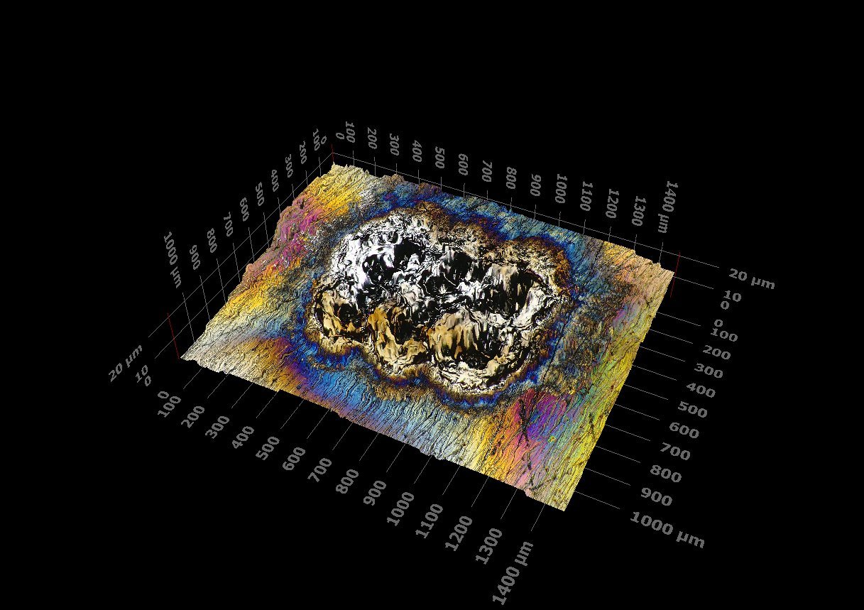
Olympus DSX1000 3D Digital Microscope
Our Olympus DSX1000 opto-digital microscope resolves surfaces in true color and full 3D, from 20x to 7000x, and measures depth, roughness, and profile without ever touching the part. It documents microstructure, fracture surfaces, corrosion damage, and wear, and it measures coating and plating thickness directly from a polished cross-section.
Laboratory
20x – 7000x
6 observation methods
Color 2D · 3D · measurement
One instrument, six ways to see a surface
The DSX1000 is a high-performance opto-digital microscope that pairs true optical resolution with digital 3D reconstruction and measurement. It is operated on-screen rather than through eyepieces, so imaging is fast, ergonomic, and repeatable. Six observation methods, brightfield, darkfield, MIX, polarization, differential interference contrast (DIC), and oblique, are switchable at a click, each one pulling out features a single mode would miss: scratches and cracks, grain and phase contrast, and the faint surface relief around a defect. The system can even recommend the best method for the sample in front of it.
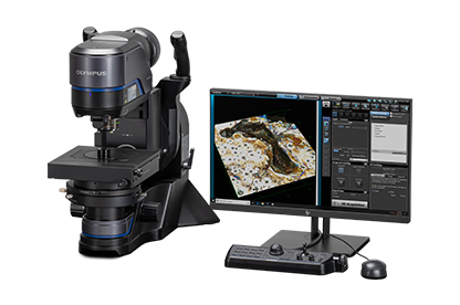
Non-contact 3D measurement
Beyond a picture, the DSX1000 reconstructs the true surface topography and measures it, step height, depth, area, volume, and surface roughness, without touching or sectioning the part. That makes it ideal for corrosion pits, wear scars, laser and LIBS burns, weld toes, and coating steps, where the number that matters is how deep, how rough, or how much material is gone.
Extended focus and high dynamic range
All-in-focus imaging keeps rough, tilted, and multi-level surfaces sharp across the entire field, and high dynamic range captures bright and dark features together, shiny metal right next to a deep black pit, in a single exposure. Automated image stitching maps large areas at high magnification without losing detail.
Where we use it
Fractography and failure analysis; LIBS and OES burn-mark evaluation; corrosion pitting depth; coating and plating thickness; weld profile and toe geometry; surface finish; inclusion and particle imaging; and dimensional checks, all documented in color and, where it counts, in measurable 3D.
Coating thickness from a cross-section
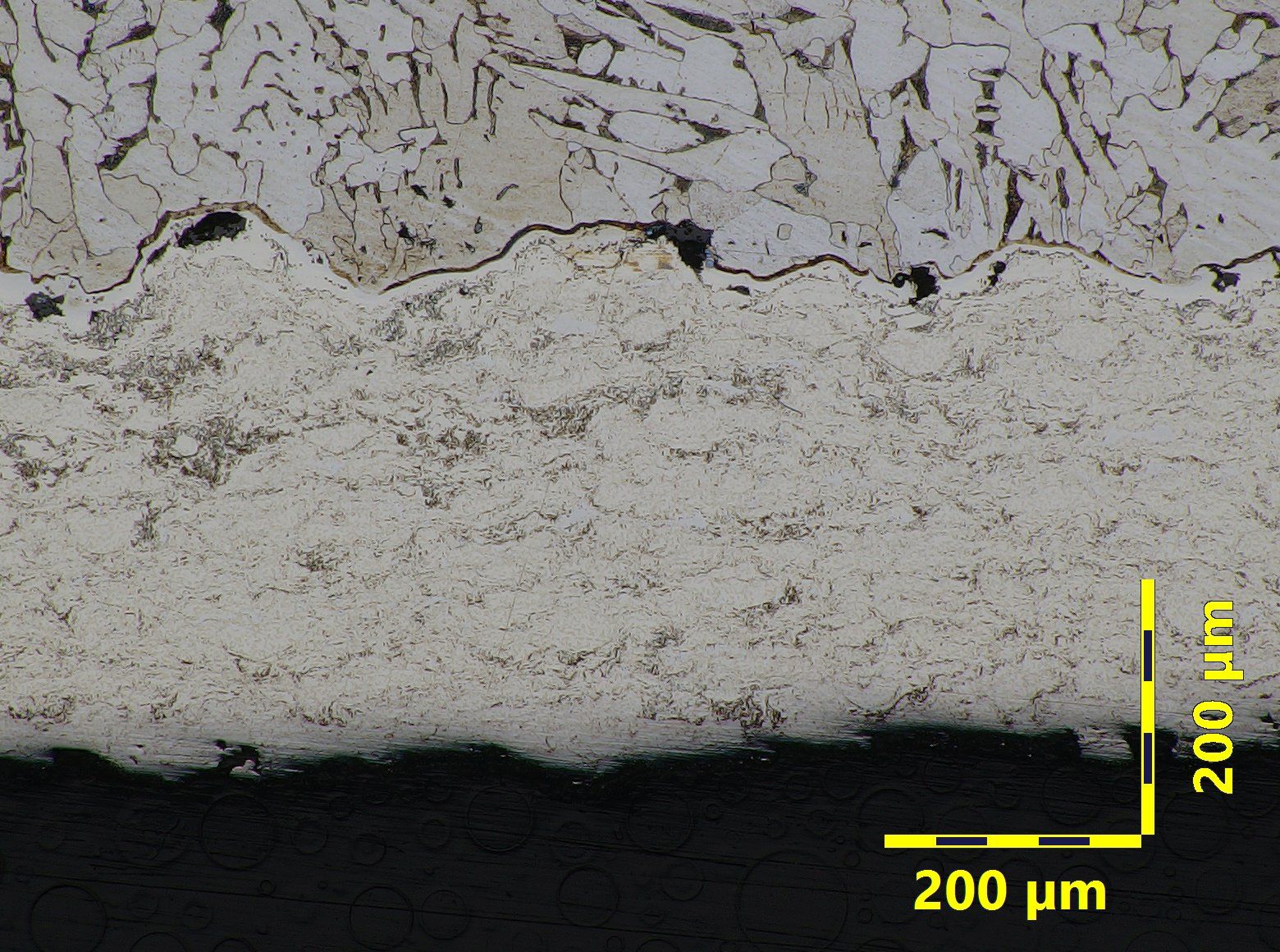
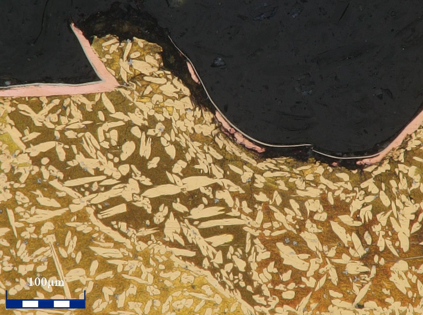
Sectioned, mounted, and polished through the coating, then measured on calibrated optics: total build-up, the individual layers of a duplex or multi-layer system, and diffusion or case depth, at as many points around the part as the specification calls for. The same section shows what a thickness gauge cannot, porosity, unfused particles, cracks, and how well the coating bonded to the substrate. See Coating Analysis for the full service.
LIBS burn, in color and in 3D
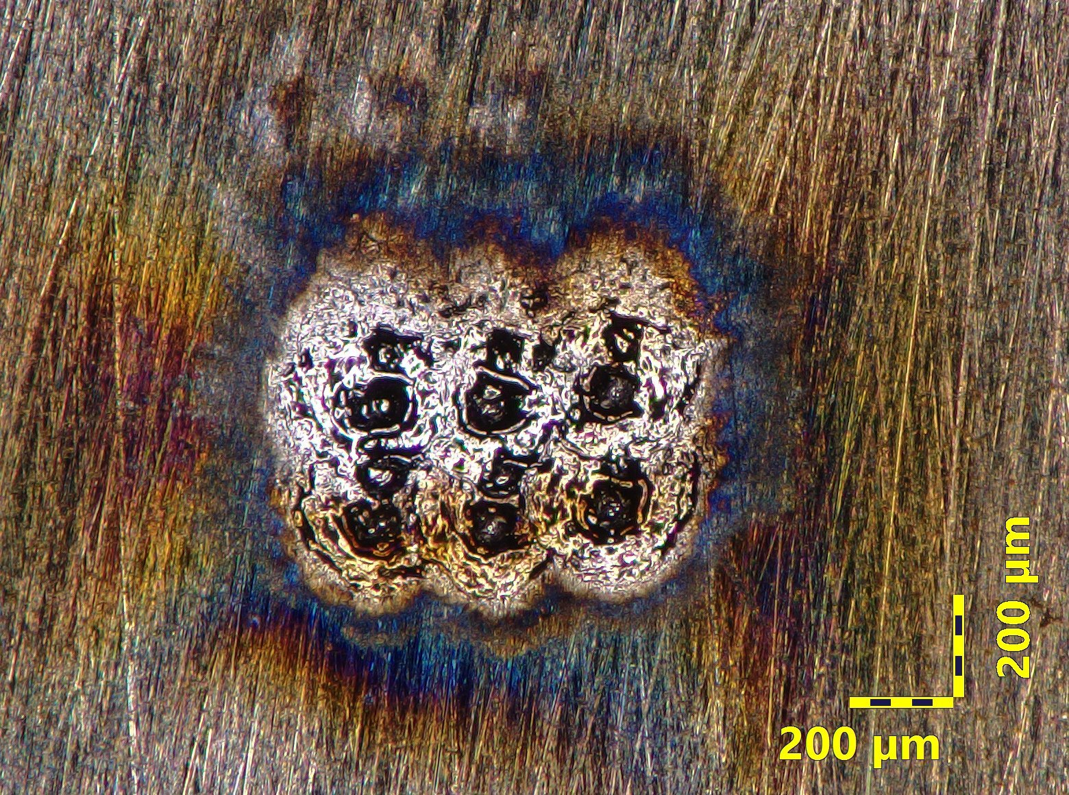
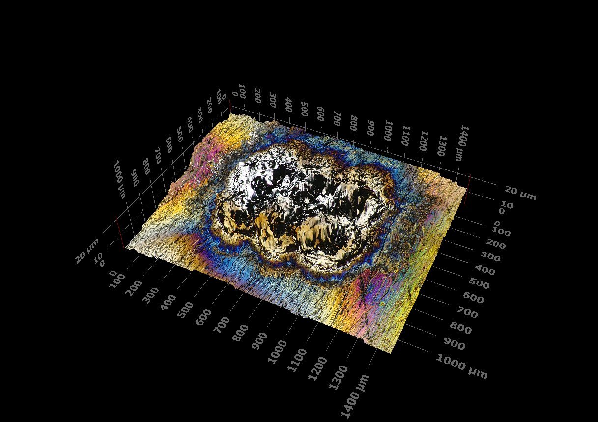
A LIBS (SciAps Z-903) burn on gray iron, imaged on the DSX1000: true-color at 160x, then reconstructed in 3D at 233x to measure the crater depth and heat-affected zone.
Often ordered together
Metallographic Examination
Sectioning, mounting, and etching to reveal grain structure and weld quality under the microscope.
View serviceMicrostructure Analysis
Grain size, phase balance, inclusions, and heat-treat condition characterized in detail.
View serviceSEM-EDS
Higher-magnification imaging with elemental micro-analysis when a case calls for it.
View serviceSee what your sample really looks like
Send us the part, the fracture, or the burn, and we will image it in true color and measurable 3D, with a report you can act on.