
SEM-EDS
Scanning Electron Microscopy paired with Energy-Dispersive X-ray Spectroscopy: high-magnification, high-depth-of-field imaging combined with elemental micro-analysis to characterize fracture surfaces, corrosion products, deposits, and inclusions.
Laboratory
SEM
EDS elemental
Fractures & deposits
What SEM-EDS does
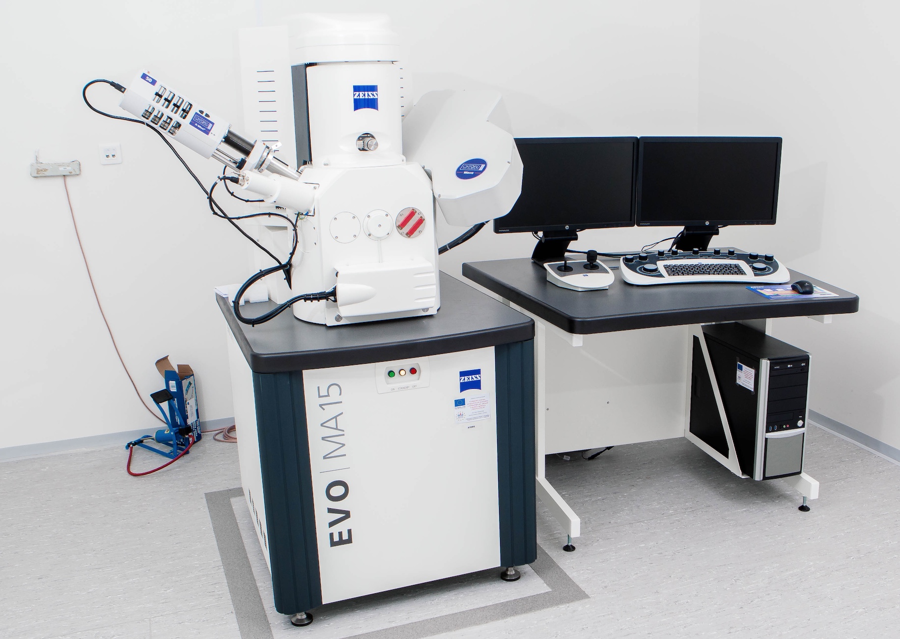
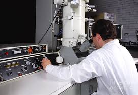
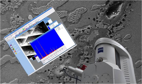
Scanning Electron Microscopy (SEM) scans a focused electron beam across a specimen to build an image from the signals it generates. Compared with light microscopy, it delivers far greater magnification and a much larger depth of field, so rough, three-dimensional surfaces stay in sharp focus across their entire relief. Paired with Energy-Dispersive X-ray Spectroscopy (EDS), the same instrument also measures the elements present at a chosen location, turning a picture of a feature into a picture and a chemistry of that feature.
Imaging that light can't reach
Because SEM keeps deep, irregular topography in focus, it excels at the surfaces that matter most in a metallurgical investigation:
- Fracture surfaces (fractography): resolving the fine features that reveal how a part broke
- Corrosion products: pits, scales, and attack morphology at the micron scale
- Deposits and scale: layered build-up on tubes, valves, and process surfaces
- Inclusions and second phases: non-metallic particles within the metal matrix
Fracture mode identification
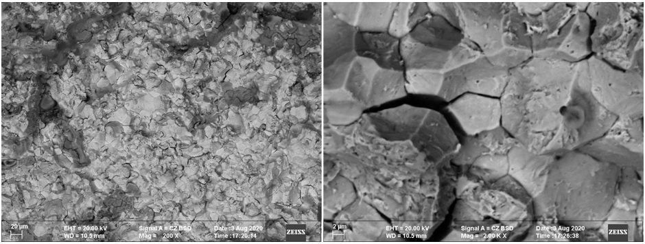
Fractography under the SEM lets us distinguish the mechanisms behind a failure by their characteristic surface signatures: fatigue striations, dimpled rupture from ductile overload, and intergranular cracking that follows grain boundaries. Reading these features is central to determining not just that a component failed, but why.
Elemental micro-analysis with EDS
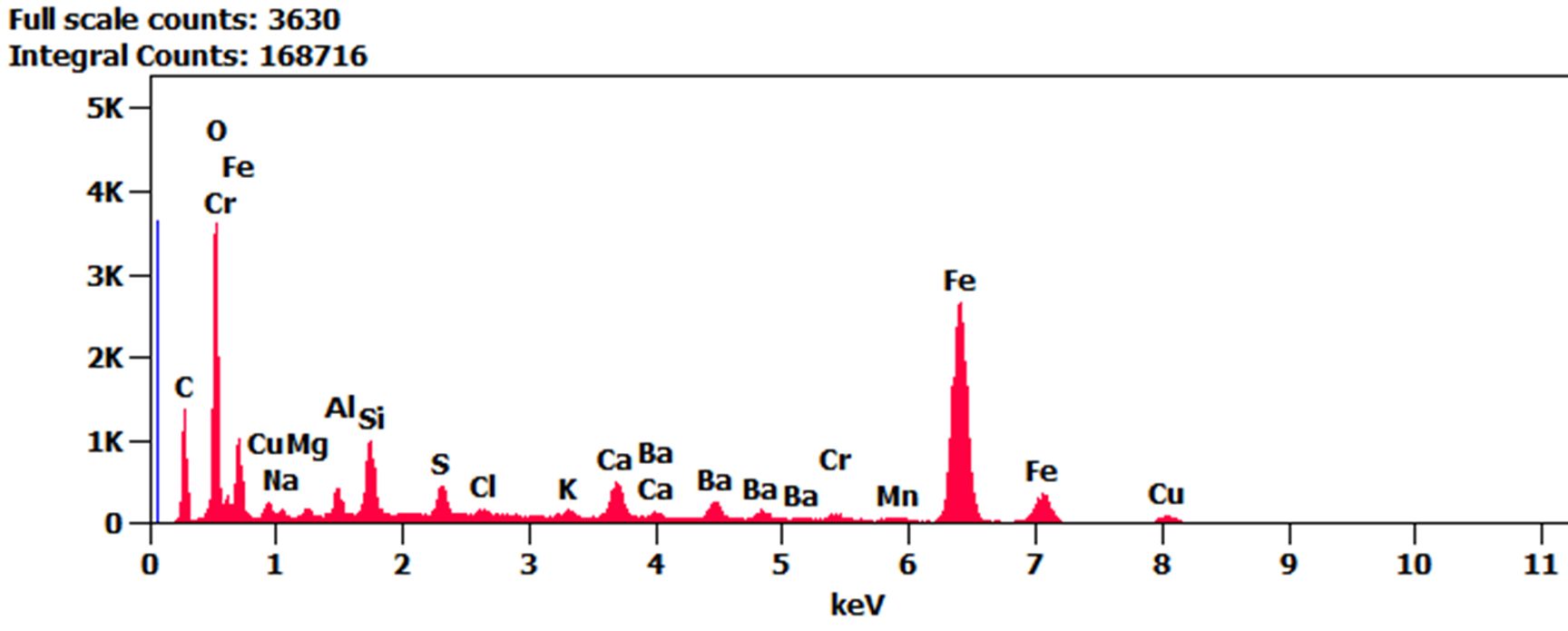
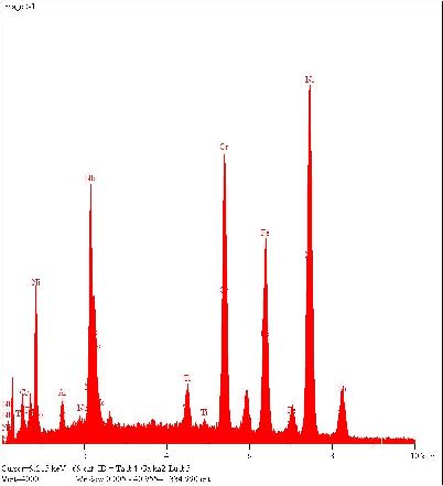
EDS collects the characteristic X-rays emitted under the electron beam to identify which elements are present. It can be run in several modes to match the question:
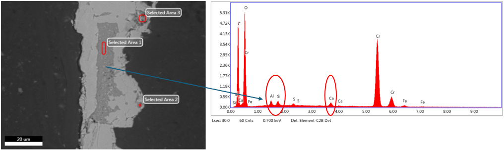
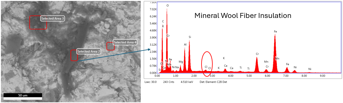
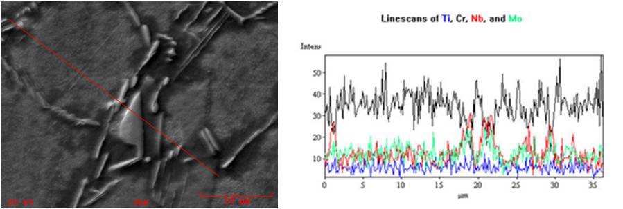
- Spot analysis: chemistry of a single feature such as an inclusion, particle, or crack origin
- Area analysis: an averaged composition across a defined region
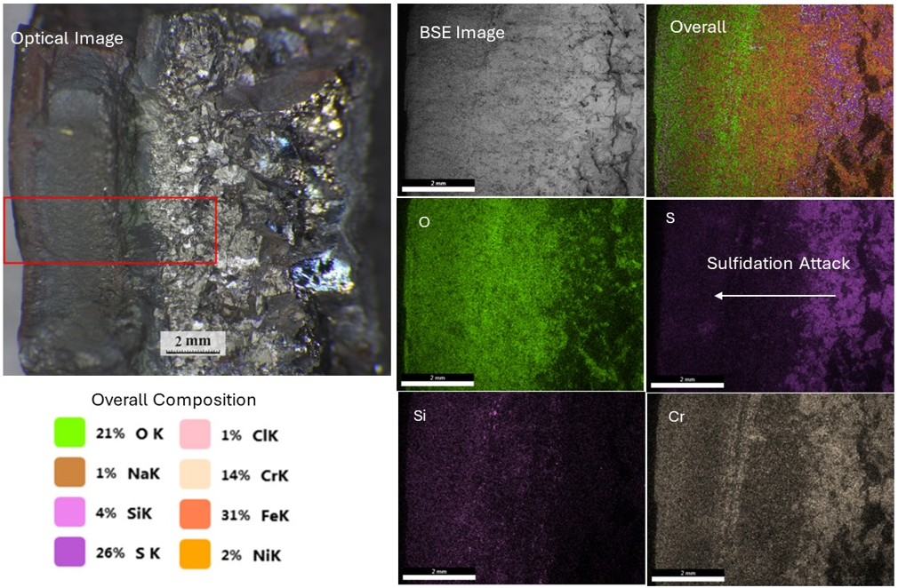
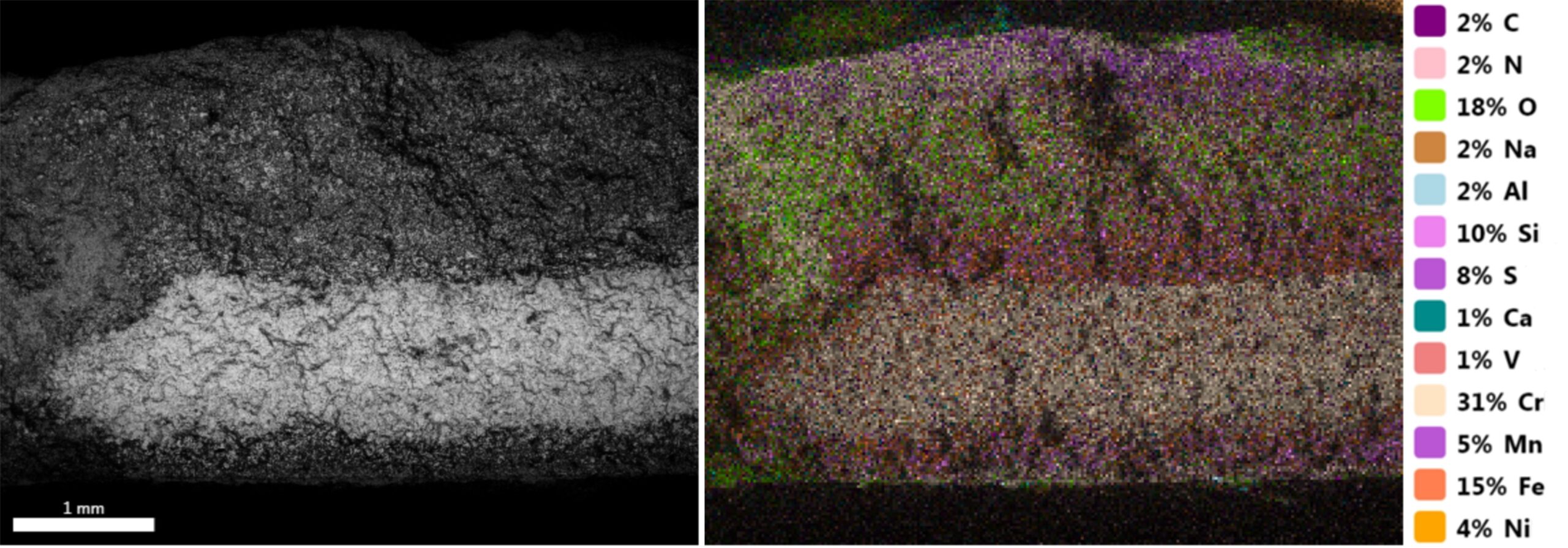
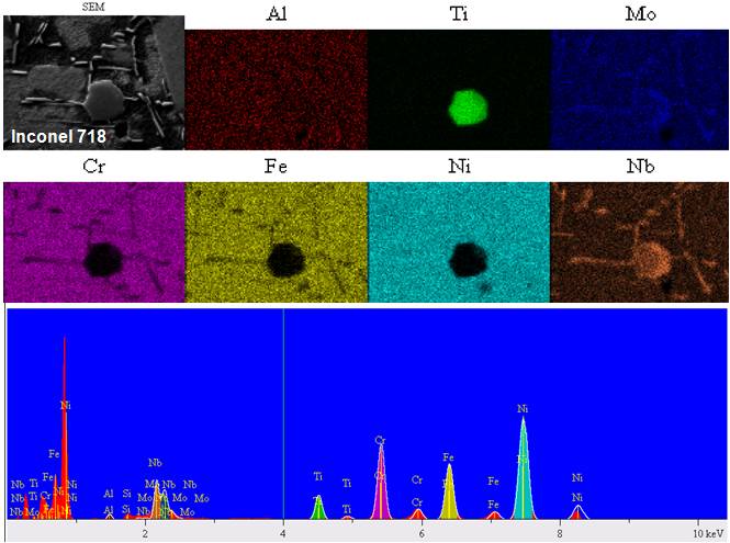
- Elemental mapping: spatial maps showing how elements are distributed across a field of view
Typical applications
SEM-EDS supports a wide range of investigations, including failure analysis and fracture mode identification, corrosion and deposit identification, weld and inclusion analysis, and contamination identification. When a component underperforms, corrodes, or fails, the combination of high-resolution imaging and on-the-spot elemental chemistry provides the evidence needed to reach a defensible conclusion.
SEM-EDS gallery
High-magnification imaging and elemental micro-analysis.
Often ordered together
Metallographic Examination
Cross-sectional study of microstructure, phases, and heat-treat condition.
View serviceFailure Analysis
Root-cause investigation built on fractographic and metallurgical evidence.
View serviceChemical Analysis
Bulk and localized composition to verify alloys and identify contaminants.
View serviceNeed to see and identify a feature?
Send us the part and the question. Our metallurgists will image the surface at high magnification and pin down its chemistry, then deliver a defensible report.